|
Poster
147 |
Where:
JMS Breakout Room (Room 745)
Speaker:
|
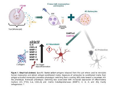
Background: Neurocysticercosis (NCC), caused by infection of the central nervous system with Taenia solium larvae, is characterised by marked neuroinflammation during parasite degeneration. Astrocytes are central regulators of inflammatory signalling and tissue homeostasis in the brain and can undergo phenotypic switching toward neurotoxic (A1) or neuroprotective (A2) states with distinct functional consequences. While astrocyte activation is a recognised feature of NCC, the mechanisms by which parasite-derived antigens influence astrocyte phenotypic responses and associated tissue remodelling processes remain poorly understood. We hypothesised that exposure to specific T. solium antigens drives astrocyte phenotypic switching and activation of tissue remodelling pathways relevant to NCC pathology.
Methods: Primary human astrocytes were exposed to conditioned media derived from primary human monocytes stimulated with defined T. solium antigens, including total parasite antigen extract (CoMTsol) and selected recombinant antigens (CoMT24, CoMGP50 and CoM8kDa). Astrocyte phenotypic switching was assessed by quantifying transcriptional markers associated with A1-like and A2-like states and measurement of inflammatory mediators secretion by ELISA (IL-6, IP-10, IL1b and TNF-a). Tissue remodelling responses were evaluated through matrix metalloproteinases (MMPs) expression and secretion, their endogenous inhibitors (TIMPs), and functional collagenase/gelatinase activity assays. To assess in vivo relevance, astrocyte phenotypes were examined in brain tissue from NCC-infected pigs using immunofluorescence and immunohistochemistry technique for astrocyte phenotype detection in viable and degenerated cysts.
Results: Astrocytes exposed to CoMTsol and CoMT24 underwent phenotypic switch to A1-like phenotype characterised by massive upregulation of c3 and ip10 genes (58.4 and 66.3-fold increase relative to unstimulated astrocytes, respectively). Pro-inflammatory responses were not significantly different from unstimulated astrocytes (p>0.9), however, elevated gene expression and secretion of MMPs were observed. Secretion of MMP-1, -3, -8, -9 and -10 were highly increased from 48 hours post-stimulation (i.e MMP-1: 1876.85 pg/ml, MMP-3: 2004.2 pg/ml and MMP-9: 3733.35 pg/ml) and collagenase/gelatinase assay confirmed activity. Tissue staining in NCC-infected pig samples also confirmed a higher population of A1-like astrocytes in the surrounding tissue of degenerated than viable cysts. On the other hand, astrocytes stimulated with CoM8kDa did not induce any specific astrocyte phenotype nor pro-inflammatory response.
Conclusion:These findings demonstrate that specific T. solium antigens drive astrocyte phenotypic switching (from A0 to A1 phenotype) and coordinate tissue remodelling responses in neurocysticercosis. This work identifies astrocytes as active contributors to parasite-driven host responses highlighting the specificity of parasite antigens to lead the astrocyte reprogramming and MMP-associated remodelling, mainly collagenases MMP-1, -3 and -10. Identification of high regulated tissue remodelling mediators could be associated with the tissue damage seen in NCC pathology and could offer new insights for the development of future host-directed therapies to control inflammation and may reduce neurological sequels in NCC.

