
|
Poster
13 |
Modulation of miRNA-721 and its host gene CUX1 in murine macrophages stimulated with LPS or infected with Leishmania |
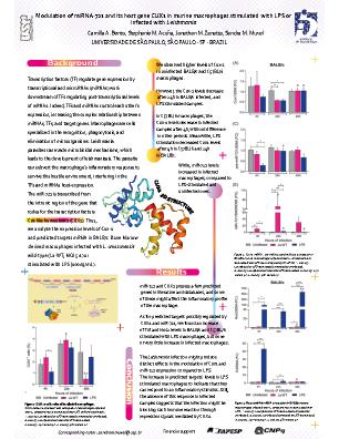
Background: Transcription factors (TF) regulate gene expression by transcriptional and microRNAs (miRNAs) work downstream of TFs regulating post-transcriptional levels of mRNAs. Indeed, TFs and miRNAs control each other’s expression, increasing the complex relationship between miRNAs, TFs, and target genes. Macrophages are cells specialized in the recognition, phagocytosis, and elimination of microorganisms. Leishmania parasites can evade microbicidal mechanisms, which leads to the development of leishmaniasis. The parasite can subvert the macrophage’s inflammatory response to survive this hostile environment, interfering in the TFs and miRNAs host-expression. The miR-721is transcribed from the intronic region of the gene that codes for the transcription factors Cut-like homeobox 1 (CUX1). Thus, we analyze the expression levels of Cux1 and predicted targets mRNA in BALB/c and C57BL/6 macrophages infected with L. amazonensis wild-type (La-WT) or stimulated with LPS. Materials and Methods: Bone-marrow-derived macrophages of BALB/c and C57BL/6 mice were infected with L. amazonensis wild-type (La-WT; MOI 5:1) or stimulated with LPS (100ng/mL) for 4, 24 e 48h (3). We performed the gene expression analysis through RT-qPCR to evaluate expression levels of Cux1 and predicted targets. Results: We observed higher levels of Cux1 in uninfected BALB/c and C57BL/6 macrophages. However, the Cux-1 levels decrease after 24h in BALB/c infected, and LPS stimulated samples. In C57BL/6 macrophages, the Cux-1 levels decrease in infected samples after 4h, without difference in other periods. Meanwhile, LPS stimulation decreased Cux1 levels after 4h in C57BL/6 and 24h in BALB/c. While, miR-721 levels increased in infected macrophages, compared to LPS-stimulated and uninfected ones. miR-721 and CUX1 possess a few predicted genes in literature and databases, and some of these might affect the inflammatory profile of the macrophage. As for predicted targets possibly regulated by CUX1 and miR-721, we found an increase of Tnf and Nos2 levels in BALB/c and C57BL/6 stimulated with LPS macrophages, but none or very little increase in infected macrophages. Conclusions: The Leishmania infection might produce distinct effects in the modulation of Cux1 and miR-721 expression compared to LPS. The increase in predicted targets’ levels in LPS stimulated macrophages to indicate that they can respond to an inflammatory stimulus. Still, the absence of this response in infected samples suggests that the infection might be blocking such immune reaction through repression signals mediated by CUX1.