
|
Poster
37 |
Characterisation of the ESAG6 and ESAG7 3’UTRs involved in the iron starvation response in Trypanosoma brucei. |
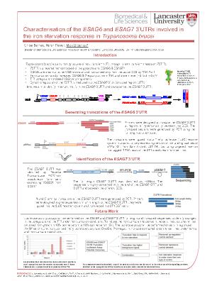
Trypanosomiasis is a neglected tropical disease caused by the protozoan parasite Trypanosoma brucei. The infection is responsible for high levels of morbidity in Sub-Saharan Africa due to the socio-economic burden of the disease (Kennedy, 2013). To be able to survive within the mammalian host, the parasite must be able to obtain iron. Mammals limit the availability of iron by sequestering it within the major serum glycoprotein transferrin, to inhibit the growth of invasive pathogens (Benz et al, 2018). T. brucei has evolved a receptor that binds transferrin with a high affinity, and therefore allows iron uptake by receptor mediated endocytosis at the flagellar pocket (Steverding et al, 1995). Under iron starvation conditions expression of the Trypanosoma brucei transferrin receptor (TbTfR) increases 5-fold, resulting in a corresponding uptake in transferrin that occurs before the intracellular stores of iron have been depleted (Mussmann et al, 2004). We have shown that this iron starvation response is mediated through the ESAG6 3’UTR (Benz et al, 2018), and in the current work we aim to identify motifs within the ESAG6 and ESAG7 3’UTRs responsible for the upregulation of the TbTfR in the iron starvation response.
The TbTfR is a heterodimeric protein encoded by two expression site associated genes, ESAG6 and ESAG7 and is anchored in the membrane by a GPI anchor on ESAG6 (Steverding, 2000). The TbTfR is evolutionarily distinct from the mammalian transferrin receptor but is structurally similar to a truncated variant surface glycoprotein (VSG) homodimer (Benz et al, 2018). T. brucei is able to sense and respond to iron starvation conditions through a post transcriptional mechanism mediated through the ESAG6 3’UTR, but the role of the ESAG7 3’UTR remains unexplored. To identify important motifs of theESAG6 and ESAG7 3’UTRs responsible for the iron starvation response we used a two-step RT-PCR reaction to identify the length of the ESAG6 and ESAG73’UTRs as 356bp and 492bp respectively. Truncations were then made of these UTRs at regions of hypervariability between the multiple BESs and ligated into a firefly luciferase reporter system. The subsequent sequences were transfected into 2T1 BSF T. brucei cells to perform luciferase activity assays under both normal and iron starvation conditions. Identified regulatory motifs could be used in combination with a selection marker to identify components in the signalling pathway to determine if any component could be used as a drug target to treat this neglected tropical disease.
References
Benz C, Urbaniak M. D, et al. (2018). Dynamic regulation of the Trypanosoma bruceitransferrin receptor in response to iron starvation is mediated via the 3’UTR. PLOS One, v.13, e020633.
Kennedy P.G.E. (2013). Clinical features, diagnosis, and treatment of human African trypanosomiasis. The Lancet Neurology, v.12, p.186-194
Mussmann R, Engstler M, et al. (2004). Factors affecting the level and localisation of the transferrin receptor in Trypanosoma brucei. The Journal of Biological Chemistry, v.279, p.40690-8.
Steverding D, Stierhof Y.D, et al, (1995). Transferrin-binding protein complex is the receptor for transferrin uptake in Trypanosoma brucei. The Journal of Cell Biology, v.131, p.1173-1182.
Steverding D. (2000). The transferrin receptor of Trypanosoma brucei. Parasitology International, v.48, p.191-8.

