Authors
A Correa da Silva2; L V Liz2; C L Pontes2; F Olmo1; A F Francisco1; F C Costa1; S Jayawardhana1; M C Taylor1; P H Stoco2; J M Kelly1; E Grisard2; 1 London School of Hygiene and Tropical Medicine, UK; 2 Universidade Federal de Santa Catarina, Brazil Discussion
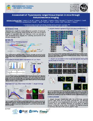
Trypanosoma rangeli is a protozoan parasite transmitted by the bite of the triatomine bugs to several vertebrate hosts. Since T. rangeli was first described by Enrique Tejera in 1920, no symptoms or physiopathological events associated with infection have been identified in mammalian hosts. T. rangeli holds an attractive biotechnological potential, due to lack of pathogenicity to mammals and phylogenetic proximity to human pathogens like Trypanosoma cruzi. To take this further, unknown features of the parasite life cycle such as tissue tropism, replication, and cell invasion need to be better understood. Here, we describe a new bioluminescence imaging tool to enhance T. rangeli research, which allows the infection to be monitored in vivo beyond the bloodstream and make possible the identification of the main infected organs. We adapted the plasmid pTRIX2 Luc::Neon, which facilitates the expression of a fluorescent:bioluminescent dual-reporter, targeting integration into the T. rangeli rDNA locus. Using an improved transfection protocol, we reduced the selection time from 3 months to 2 weeks. The resulting plasmid named pTRIXrang can be stably integrated into T. rangeli strains from different genetic backgrounds (SC58 and Choachí), resulting in high level of expression in the absence of the selective drug in >90% of the parasite population. Metacyclogenesis in Rhodnius prolixus and parasitemia in BALB/c mice were assessed. No alterations in infection kinetics or parasite numbers were found in these hosts when infected by wild type and genetically modified reporter strains – Choachí (p=0.33) and SC58 (p=0.19). In vivo bioluminescence revealed that by 13 days post-infection in BALB/c mice, the parasites load had decreased below the detection threshold. During the early acute phase of infection, the spleen and lungs were the organs with the highest parasite burden. Additionally, the prostatic lobe and gonadal adipose tissue of male mice were also sites of parasite sequestration. Non-classical motionless round-shaped parasites could be detected in the bloodstream. The replicative state of the parasite is under current investigation. Therefore, we describe the first integrative T. rangeli specific plasmid and the resulting modified parasite lines that express high levels of a luciferase:mNeonGreen dual-reporter, that does not alter the parasite biology in vitro and in vivo, allowing downstream applications. As illustrated, this has considerable potential to boost the host-parasite studies and shed light on the T. rangeli life cycle in mammalian hosts.